


Excerpted from
BEYOND MAMMOGRAPHY
by Len Saputo, MD,
Health Medicine Institute
www.healthmedicinecenter.net
 Breast thermography has been available in clinical practice since the 1960s. Initially, physicians were very excited when they learned that breast cancers emit more infrared heat than normal healthy tissues, and that they could be detected using infrared scanners. However, this technology was brought into practice prematurely—before clinical trials were completed, and before sufficient information about other health conditions that also emitted large amounts of infrared light were understood.
Unfortunately, this resulted in many women having breast surgeries that did not have breast cancer. Eventually, the high rate of unneeded surgeries led to the rejection of infrared breast imaging in the United States, with the entire technology being sidelined by mainstream medical practice for several decades.
Since the 1970s, however, clinical research has continued, especially in Canada and France where this technology is considered more mainstream. More than 800 research papers have been published on the subject of breast thermography, and a research databank on more than 300,000 women who have been tested with infrared breast imaging now exists.
In addition, major advances in infrared imaging technology have been achieved that improve the sensitivity to 0.05 degrees centigrade, which makes identifying breast cancer much easier and more reliable. The combination of improved technology and scientific clinical research is sparking the return of breast thermography into clinical practice today.
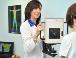
How Breast Thermograms Work
Breast thermography measures differences in infrared heat emission from normal breast tissue, benign breast abnormalities—such as fibrocystic disease, cysts, infections and benign tumors—and from breast cancers. It does this with a high degree of sensitivity and accuracy. Breast thermography is a non-invasive measurement of the physiology of breast tissue. This technology is not meant to replace mammography or other diagnostic tests presently used in clinical practice that measure anatomical abnormalities in breast tissue. While breast cancer can only be diagnosed by tissue biopsy, breast thermography safely eliminates the need for most unnecessary biopsies as well as their associated high cost and emotional suffering, and it does so years sooner than any other test in modern medicine.
Modern infrared scanners have a thermal sensitivity of 0.05 degrees Centigrade. Because tumor tissue does not have an intact sympathetic nervous system, it cannot regulate heat loss. When the breast is cooled with small fans in a room kept at 68 degrees Fahrenheit, blood vessels of normal tissue respond by constricting to conserve heat while tumor tissue remains hot. Thus, tumors emit more heat than their surrounding tissues and are usually easily detected by heat-sensing infrared scanners.
Over time, cancerous tissues stay hot or become even hotter—they do not cool down. In sharp contrast, however, other possible conditions such as fibrocystic breasts, infections, and other benign disorders cool down as they resolve.
Breast thermograms have highly specific thermal patterns in each individual woman. They provide a unique “thermal signature” that remains constant over years unless there is a change in an underlying condition. Thus, over time, it is possible to differentiate between cancers and benign conditions. Based on this ability to more accurately detect cancers over time, it becomes important to have a benchmark early on in a woman’s life. For this reason, women should have breast thermography performed beginning at age 25.
Thermograms are graded with a system much like pap smears with grades 1-5. Th1 and Th2 are normal, Th3 is moderately abnormal, and Th4 and Th5 are severely abnormal and require careful follow-up because many of them are caused by cancer. Of significance, one recent study documented that women with Th1 and Th2 scores can be reassured with a 99 percent level of confidence that they do not have breast cancer. (16)
Clinical Research Supporting Breast Thermography
At least five important studies published between 1980 and 2003 document that breast thermal imaging is a major advancement in identifying breast cancers not only with greater sensitivity and specificity, but also years earlier than with any other scientifically tested medical technology.
These scientific studies include:
- Cancer, 1980, Volume 56, 45-51. (17) Fifty eight thousand patients with breast complaints were examined between 1965 and 1977. Twelve hundred and forty five patients with abnormal Th3 mammotherms had normal breasts by mammography, ultrasound, physical exam, and biopsy. Thirty-eight percent of women with normal breasts and 44 percent of those with mastopathy developed biopsy proven breast cancer within five years. Ninety percent of patients with Th4 or 5 had diagnosis of cancer made on their first visit.
- Biomedical Thermology, 1982, 279-301, Alan Liss, Inc, NY. Michel Gautherie, MD, followed 10,834 women over 2 to 10 years by clinical examination, mammography and thermography. (15) The study followed 387 people with normal breast examinations and mammograms but Th3 thermographic scores for an average of less than three years. In those without symptoms, 33 percent developed cancer. In those with cystic mastitis, cancer developed in 41 percent. These were predominately women between 30 to 45 years of age where breast cancer is the leading cause of death.
- Thermology, 1986, Volume 1, 170-73. (18) The effectiveness of mammography, clinical palpation, and thermography were compared in the detection of breast cancer. Thermography had the best reliability, but the best results were found when all three were used together.
- The Breast Journal, Volume 4, 1998, 245-51. (19) Keyserlingk et al documented 85 percent sensitivity in diagnosing breast cancer using clinical examination and mammography together. This increased to 98 percent when breast thermography was added.
- American Journal of Radiology, January 2003, 263-69. (16) The journal reported that thermography has 99 percent sensitivity in identifying breast cancer with single examinations and limited views. Thus, a negative thermogram (Th1 or Th2) in this setting is powerful evidence that cancer is not present.
Important Highlights from Breast Thermography Studies
- Advances in infrared technology combined with data on 300,000 women with mammotherms document that breast thermography is highly sensitive and accurate. Today, this means that more than 95 percent of breast cancers can be identified, and that this is done with 90 percent accuracy. In women under the age of 50, where there is the most devastating loss of life from breast cancer, mammography, MRIs and PET scans cannot come close to matching the combined sensitivity and specificity (accuracy) of breast thermography.
- Breast thermography involves no radiation exposure or breast compression, is easy to do, is done in a private setting, and is affordable.
- The FDA approved breast thermography for breast cancer risk assessment in 1982.
- It is important to begin breast cancer screening long before age 40. It should begin at age 25 in order to identify young women who are already developing breast cancer since it takes approximately 15 years for a breast cancer to form and lead to death. Further, young women with dense breast tissue are the most difficult to evaluate using breast palpation, mammography, and ultrasound examinations, yet their significantly higher risk of developing breast cancer can be accurately detected with breast thermography.
- Mainstream procedures are not approved for breast cancer screening in women under age 40—it is widely known and accepted that they miss too many cancers and lead to too many false positive findings that result in far too many needless breast biopsies.
Conclusion
There is an abundance of scientific evidence supporting that breast thermography is the most sensitive and accurate way to identify women with breast cancer, especially in women under the age of 55, where it causes the most devastating loss of life. For women over 55, breast thermography is an important adjunct to clinical breast examination and mammography, as this combination has been documented to increase identification of breast cancers to 98 percent.
Because of its low cost and high degree of sensitivity and accuracy, all women who want to be screened for breast cancer should begin having breast thermograms beginning at age 25. Clearly, there are situations that warrant the use of other modalities such as mammography, ultrasound, MRI, PET scanning, nipple aspirations, or biopsy, and these valuable tools should continue to be used in clinical practice along with breast thermography.
Many new technologies are on the horizon that may become mainstream in the near future. With the advent of highly sophisticated genetic technology, new proteins are constantly being discovered that offer promise as markers of early breast cancer. (20) Recently published reports also suggest that MRI technology may be blended with spectrophotometric measurements that could diagnose breast cancer without even doing a biopsy. (21)
The practice of medicine, just like everything in life, is in constant evolution—there is no guarantee that what is in the mainstream today will be here tomorrow. Yet, the advancement of all fields of endeavor often moves slowly and cautiously, sometimes at the expense of human life. We must remain open and alert as new, exciting, and safe strategies emerge, especially in situations where there is such a pressing need for new approaches.
REFERENCES
- Elliott, V S. Mammography debate: Who should get screened and when? American Medical News, an AMA publication. Volume 10, number 42, pages 35-37, November 10, 2003. www.amednews.com.
- Kerlikowske, K. Use of mammograms in older women questionable. JAMA. December 10, 2003.
- Time Magazine, April 28, 2003. Cover story: The No. 1 Killer of Women.
- SEER, National Cancer Institute: Chances of developing breast cancer at a given age.
- de Sanjose S, et al. Prevalence of BRCA1 and BRCA2 germline mutations in young breast cancer patients: a population-based study. Int J Cancer 2003; 106 (4): 588-93.
- Furstenberger et al. Insulin like growth factors mediate breast cancer growth and proliferation. Onkologie, 2003. Volume 26, number 3, pages 290-94.
- Baker L. Breast cancer detection demonstration project: Five year summary report. Cancer, 1982, volume 32, pages 194-225.
- Sickles EA. Breast masses: mammographic evaluation. Radiology 1989. Pages 173-303.
- Fletcher, S W, and Elmore, J G. Mammographic Screening for Breast Cancer. New England Journal of Medicine. Volume 348, no. 17, pages 1672-80. April 24, 2003.
- Ostbye, T. Elderly women over-screened for cancers with little measurable benefit. Annals of Family Practice. November/December issue, 2003.
- Pisano, E. Digital Mammography Offers Better Breast Cancer Diagnoses. Presented at the Radiologic Society of North America annual meeting, December 2003. Research conducted at University of North Carolina School of Medicine. etpisano@med.unc.edu.
- Freidrich M. MRI of the breast: State of the art. European Radiology, 1998. Volume 8, pages 707-725.
- Avril N, Rose CA, Schelling M, et al. Breast imaging with positron emission tomography and fluorine-18 flourodeoxyglucose: use and limitations. Journal of Clinical Oncology, 2000. Volume 18, pages 3495-3502.
- Avril N. Discussions in PET Imaging 2003. CMP Healthcare Media, DPI no. 621, PET and Breast Cancer.
- Gautherie, M, Haehnel, P, Walter, J p, Keith, L. Long-Term Assessment of Breast Cancer Risk by Liquid-Crystal Thermal Imaging. Biomedical Thermology, pages 279-301. 1982 Alan R. Liss, Incl, 150 Fifth Avenue, New York, NY 10011.
- Parisky, Y R, et al. Efficacy of Computerized Infrared Imaging Analysis to Evaluate Mammographically Suspicious Lesions. American Journal of Roentgenology, January 2003, 263-69.
- Gautherie, M, and Gros, C M. Breast Thermography and Cancer Risk Prediction. Cancer, 1980, volume 56, 45-51.
- Nyirjesy, M D, et al. Clinical Evaluation, Mammography and Thermography in the Diagnosis of Breast Carcinoma. Thermology, 1986, volume 1, 170-73.
- Keyserlingk, M D, et al. Infrared Imaging of the Breast: Initial Reappraisal Using High-Resolution Digital Technology in 100 successive cases of Stage I and II Breast Cancer. The Breast Journal, volume 4, 1998, 245-51.
- Zangar, R. Breast Cancer Research and Treatment. July 3, 2003.
Bolan, P. In vivo quantification of choline compounds in the breast with 1H MR spectroscopy. Magnetic Resonance in Medicine. Volume 50, Issue 6, Date: December 2003, Pages: 1134-1143.